Nanopartz Fluorophore-Labeled Gold Nanoparticles for Miniflux Microscopy: Achieving Ultimate Resolution
Nanopartz fluorophore-labeled gold nanoparticles are revolutionizing the field of fluorescence microscopy, particularly with Miniflux Microscopy, a technique designed to push the boundaries of resolution beyond traditional limits. These advanced nanoparticles enable researchers to capture high-definition images at unprecedented clarity and scale, making them an invaluable tool in scientific imaging and research.
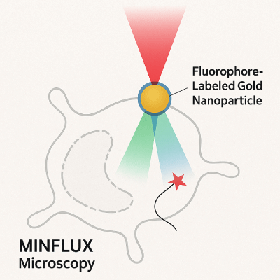
A visual representation of Nanopartz fluorophore-labeled gold nanoparticles in use with Miniflux Microscopy |
|---|
Properties of Nanopartz Fluorophore-Labeled Gold Nanoparticles
Nanopartz fluorophore-labeled gold nanoparticles are distinguished by their unique combination of gold’s biocompatibility with the light-emitting properties of attached fluorophores. This combination results in:
- Gold Core: Gold nanoparticles offer exceptional optical and chemical stability. Their unique interaction with light enhances imaging capabilities, especially at the nanoscale.
- Fluorophore Labeling: The fluorophores attached to the nanoparticles emit specific wavelengths of light when excited by a laser, enabling bright and sharp fluorescence signals.
- Size Control: Nanopartz offers precise size control ranging from 1 nm to 100 nm, ensuring customizability for different applications in microscopy and research.
Applications of Nanopartz Fluorophore-Labeled Gold Nanoparticles
-
Super-Resolution Imaging: Nanopartz fluorophore-labeled gold nanoparticles allow researchers to break the diffraction limit in Miniflux Microscopy, achieving resolutions below 10 nm. This makes it possible to observe molecular structures in unprecedented detail.
-
Cellular and Molecular Imaging: These nanoparticles are used to label specific proteins, organelles, or nucleic acids, providing high-contrast images in biological samples. This application is critical for studying disease mechanisms and cell biology.
-
Tracking Molecular Dynamics: The unique optical properties of the labeled nanoparticles make them ideal for tracking fast-moving molecules and proteins inside living cells in real-time.
How They Work: Enhancing Miniflux Microscopy
Miniflux Microscopy, which focuses on maximizing the efficiency of fluorescence emission and detection, benefits greatly from the use of fluorophore-labeled gold nanoparticles. Here’s how they contribute:
-
Enhanced Signal: The gold core amplifies the fluorescence signal by increasing the local electromagnetic field around the fluorophore, resulting in a brighter signal.
-
Stability and Sensitivity: Gold nanoparticles provide unmatched chemical stability, ensuring that fluorescence signals remain strong over extended imaging periods. This is crucial for capturing high-resolution images with minimal background noise.
-
Precision Targeting: Nanopartz’s proprietary technology ensures that the fluorophores are densely packed around the gold nanoparticles, allowing precise targeting of biological structures.
Workflow: Utilizing Nanopartz Nanoparticles in Miniflux Microscopy
-
Sample Preparation: The sample is prepared by conjugating Nanopartz fluorophore-labeled gold nanoparticles to target molecules (e.g., proteins, nucleic acids).
-
Microscope Setup: The sample is placed under a Miniflux Microscope, where a high-powered laser excites the fluorophores attached to the nanoparticles.
-
Fluorescence Detection: The excited fluorophores emit light, which is captured by the microscope's detector. Thanks to the gold core, the signal is amplified, leading to a highly detailed and bright image.
-
Image Processing: The data from the fluorescence is processed to generate super-resolution images, revealing structures that are otherwise beyond the diffraction limit of conventional microscopes.
Advantages of Nanopartz Fluorophore-Labeled Gold Nanoparticles
-
Higher Resolution: Nanopartz nanoparticles are instrumental in achieving the ultimate resolution in fluorescence microscopy, making them ideal for cutting-edge research.
-
Biocompatibility: The gold nanoparticles are biocompatible, ensuring safe use in live-cell imaging without compromising cellular functions.
-
Increased Sensitivity: Fluorophore labeling enhances detection sensitivity, allowing for the observation of even the most minute biological processes.
-
Long-Lasting Stability: Nanopartz particles resist photobleaching, ensuring consistent imaging quality over long periods.
Conclusion
Nanopartz fluorophore-labeled gold nanoparticles are a game-changer in fluorescence microscopy, especially when paired with Miniflux Microscopy. Their ability to push the resolution limits and provide stable, bright fluorescence signals is key to advancing biological research. Whether for cellular imaging or tracking molecular dynamics, these nanoparticles offer unparalleled performance, making them indispensable in modern scientific endeavors.
References
- Nanopartz. “Fluorophore Labeled Gold Nanoparticles for Imaging.” Nanopartz.com, accessed September 2024.
- Grabner et al (2022) Resolving the molecular architecture of the photoreceptor active zone with 3-D Microflux https://www.science.org/doi/full/10.1126/sciadv.abl7560.
Go here to purchase Nanopartz Gold Nanoparticles for Minflux microscopy applications


