Photothermal Microscopy Using Nanopartz Nanoparticles
Introduction to Photothermal Microscopy
Photothermal microscopy is an advanced imaging technique leveraging the photothermal effect to study biological systems at the cellular and molecular levels. By utilizing gold nanoparticles from Nanopartz™, researchers can achieve precise thermal manipulation of cells and tissues, followed by high-resolution imaging to assess biological responses. This dual capability makes photothermal microscopy a critical tool in applications like cancer research, drug delivery, and diagnostics.
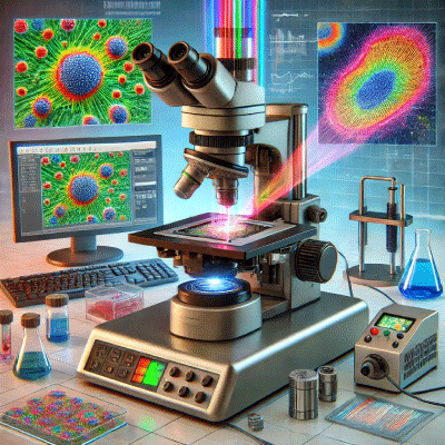
Illustration of a photothermal microscopy setup. It includes a high-resolution microscope, a laser light source connected via fiber optics, a sample stage with a biological sample, and a detector. |
Nanopartz Products for Photothermal Microscopy
Nanopartz is a leader in the field of gold nanoparticle technology, offering products specifically designed for photothermal microscopy and imaging. Their nanoparticles are recognized for their:
- Enhanced Photothermal Efficiency: Ideal for precise thermal modulation.
- Custom Functionalization: Tailored to bind specific biomolecules or target cellular receptors.
- Exceptional Optical and Thermal Properties: High scattering efficiency and stability.
Key Features
- Precision Engineering: Nanopartz provides custom conjugated gold nanorods with narrow size distributions, ensuring consistent thermal and optical properties.
- Targeted Applications: Surface modifications enable nanoparticles to target specific biomarkers or receptors, facilitating advanced imaging and localized therapeutic applications.
- Biocompatibility: Coatings minimize cytotoxicity, making them suitable for in vivo and in vitro studies.
Applications of Photothermal Microscopy with Nanopartz Products
-
Cancer Detection and Therapy
- Research Insight: Studies have demonstrated the use of Nanopartz gold nanoparticles in detecting and thermally ablating cancer cells with high precision (Nedosekin et al., 2013; Manikandan et al., 2013).
- Outcome: These nanoparticles enable simultaneous photothermal therapy and imaging, offering real-time monitoring of tumor responses.
- Drug Delivery Studies
- Nanoparticles functionalized with therapeutic agents allow for triggered drug release upon photothermal activation (Park et al., 2010).
- This capability ensures that drugs are delivered directly to the targeted site, reducing systemic toxicity.
- Live-Cell Imaging
- Nanopartz’s conjugated gold nanorods have been used to perform multicolor photothermal confocal microscopy, enabling visualization of dynamic processes in live cells (Nedosekin et al., 2015).
- Quantitative Histology
- Photothermal multispectral imaging with Nanopartz nanoparticles allows for detailed analysis of nanoparticle distribution in tissues and micrometastasis (Nedosekin et al., 2010).
- Thermal Diffusivity Measurements
- Nanopartz products facilitate precise thermal diffusivity measurements at the single-particle level, aiding in the characterization of various materials (Heber et al., 2017).
Photothermal microscopy is a versatile imaging technique that has found applications across a variety of fields due to its sensitivity and specificity. Here are some key applications:
1. Biomedical Research
- Cancer Detection and Therapy:
- Detect circulating tumor cells (CTCs) using gold nanoparticles as contrast agents.
- Monitor the efficacy of photothermal therapy in real-time by visualizing thermal stress on cancer cells.
- Drug Delivery Studies:
- Visualize and track nanoparticles delivering drugs to targeted cells or tissues.
- Analyze photothermal-triggered drug release mechanisms.
- Live-Cell Imaging:
- Observe intracellular processes without invasive fluorescent markers, preserving cellular integrity.
2. Material Science
- Thermal Conductivity Studies:
- Measure thermal diffusivity of materials at the nanoscale.
- Study heat transfer properties in composites and nanostructures.
- Characterization of Nanoparticles:
- Quantify the optical and thermal properties of nanoparticles.
- Assess nanoparticle aggregation and stability in various environments.
3. Environmental Science
- Pollutant Detection:
- Detect and analyze microplastics or pollutants in water using functionalized nanoparticles.
- Study the thermal properties of aerosols and their environmental impact.
4. Advanced Imaging Techniques
- Photoacoustic Imaging Integration:
- Combine photothermal and photoacoustic techniques to achieve multimodal imaging for deeper tissue penetration and enhanced contrast.
- Dynamic Imaging:
- Use photothermal microscopy to visualize dynamic interactions, such as nanoparticle uptake by cells or molecular interactions in real-time.
5. Diagnostics
- Pathogen Detection:
- Identify bacteria, viruses, or other pathogens using nanoparticles conjugated with specific antibodies.
- Monitor infection progression and treatment efficacy in real-time.
- Non-Invasive Blood Analysis:
- Study hemoglobin and other biomolecules with photothermal contrast, aiding in diagnostic applications.
6. Cellular and Molecular Biology
- Thermal Stress Studies:
- Analyze cellular responses to localized heating, such as apoptosis, necrosis, or heat shock protein activation.
- Investigate molecular changes induced by thermal effects at the single-cell level.
- Protein and Enzyme Studies:
- Study the thermodynamics of protein interactions and enzyme activities.
7. Nanomedicine Development
- Theranostics:
- Combine therapy and diagnostics in a single platform to deliver treatments and monitor outcomes simultaneously.
- Multimodal Imaging Platforms:
- Develop hybrid techniques that integrate photothermal microscopy with fluorescence or other imaging modalities.
8. Energy Research
- Solar Absorber Studies:
- Investigate the efficiency of nanoscale solar absorbers by analyzing their thermal properties.
- Thermoplasmonic Applications:
- Optimize materials for applications like photothermal energy conversion and storage.
Photothermal microscopy’s ability to deliver label-free, high-resolution, and dynamic imaging makes it indispensable in research and applied sciences.
Advantages of Nanopartz Nanoparticles
- Multimodal Functionality: Suitable for photothermal, photoacoustic, and fluorescence imaging.
- Enhanced Signal Strength: High resonance efficiency leads to sharper, more detailed images.
- Custom Solutions: Tailored nanoparticles to meet specific research requirements.
Research Highlights
-
Multicolor Photothermal Confocal Microscopy
- Study: Nedosekin et al., 2015.
- Key Insight: Nanopartz custom gold nanorods were used to image live cells in multicolor photothermal microscopy.
- Impact: This innovation enables the visualization of cellular interactions in real-time with unparalleled resolution.
- Photothermal Image Cytometry
- Study: Nedosekin et al., 2010.
- Key Insight: Nanopartz nanoparticles enhanced imaging in intact tissues, enabling the detection of micrometastases.
- Impact: Facilitates the early detection of cancer metastasis.
- Photoacoustic Detection of Tumor Cells
- Study: Galanzha et al., 2015.
- Key Insight: Nanopartz products were instrumental in detecting circulating tumor cells in cerebrospinal fluid via photothermal microscopy.
- Impact: A breakthrough in non-invasive cancer diagnostics.
Technical Specifications
| Property | Description |
|---|---|
| Particle Type | Gold nanorods, spherical gold nanoparticles |
| Sizes Available | 10 nm to 100 nm |
| Surface Functionalization | Antibody, peptide, PEG |
| Wavelength Compatibility | Tunable to 740 nm and beyond |
| Biocompatibility | Low cytotoxicity coatings for safe biological use |
| Applications | Cancer detection, drug delivery, thermal diffusivity |
References:
- Nedosekin, D. A., et al. "Photothermal confocal multicolor microscopy of nanoparticles and nanodrugs in live cells." Drug Metabolism and Disposition, 2015. Taylor & Francis.
-
Nedosekin, D. A., et al. "Photothermal multispectral image cytometry for quantitative histology of nanoparticles and micrometastasis in intact tissues." Cytometry Part A, 2010. Wiley Online Library.
-
Galanzha, E. I., et al. "Photoacoustic and photothermal cytometry using photoswitchable proteins and nanoparticles with ultrasharp resonances." Journal of Biomedical Optics, 2015. Wiley Online Library.
-
Heber, A., et al. "Thermal diffusivities studied by single-particle photothermal deflection microscopy." ACS Photonics, 2017. ACS Publications.
-
Park, J. H., et al. "Cooperative nanoparticles for tumor detection and photothermally triggered drug delivery." Pharmaceutical Research, 2010. NCBI.
Go here to purchase Functionalized Gold Nanoparticles
Go here to purchase Gold Nanoparticles for Cell Uptake
Go here to purchase Gold Nanoparticles for Nuclei Uptake


