
Using Gold Nanoparticles in X-Ray Imaging to Outperform Blood Tests in Detecting Kidney Disease
Kidney disease detection has traditionally relied on blood tests to assess biomarkers like creatinine and blood urea nitrogen (BUN). While these methods are widely used, they are often limited by their sensitivity, providing results only after significant kidney damage has already occurred. The use of gold nanoparticles (AuNPs) in X-ray imaging offers a groundbreaking alternative that can outperform these blood tests by detecting kidney dysfunction earlier and with greater accuracy.
Gold nanoparticles provide enhanced imaging contrast and can be engineered to target specific tissues. This makes them particularly useful for non-invasive imaging techniques like X-ray computed tomography (CT), enabling researchers and clinicians to visualize kidney structures in much finer detail.
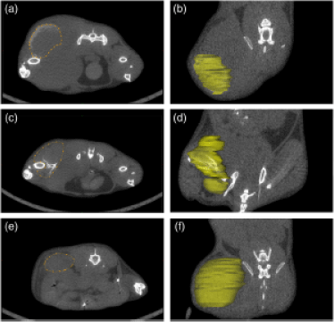
Image comparing gold nanoparticle-enhanced X-ray imaging with traditional blood tests for detecting kidney disease. This visual will complement your web page, helping to illustrate how AuNPs provide superior imaging detail. |

How Gold Nanoparticles Work in X-Ray Imaging
Gold nanoparticles have unique properties that make them ideal for medical imaging:
- High atomic number: Gold's high atomic number increases X-ray absorption, making gold nanoparticles highly visible in X-ray and CT scans.
- Biocompatibility: AuNPs are well-tolerated in biological systems and can be functionalized to target specific cells or tissues, including the kidneys.
- Targeted delivery: Functionalized AuNPs can be designed to accumulate in specific regions of the kidney, allowing for precise imaging of abnormalities like fibrosis or early-stage kidney disease.
When administered intravenously, gold nanoparticles enhance the contrast in X-ray imaging, allowing for earlier detection of kidney disease compared to traditional blood tests. While blood tests measure indirect markers of kidney function, gold nanoparticle-enhanced X-ray imaging provides a direct view of the kidney’s condition, enabling earlier intervention.
Advantages of Gold Nanoparticles in Kidney Disease Detection
-
Higher Sensitivity: X-ray imaging using gold nanoparticles can detect subtle changes in kidney tissue structure, such as early-stage fibrosis or inflammation, that blood tests cannot identify until later stages.
-
Non-invasive and Direct Visualization: Unlike blood tests that infer kidney function indirectly, AuNP-enhanced X-ray imaging allows clinicians to directly visualize the kidneys, giving a clearer understanding of disease progression.
-
Functional Imaging: AuNPs can be functionalized to bind to specific cellular markers in kidney tissue, enabling functional imaging that highlights areas of early damage or disease that would otherwise go unnoticed.
-
Faster Diagnosis: Early and accurate detection of kidney disease with gold nanoparticles means that treatment can be initiated sooner, potentially slowing or preventing disease progression.
X-Ray Imaging with Gold Nanoparticles vs. Blood Tests
Aspect Gold Nanoparticles in X-Ray Imaging Traditional Blood Tests Detection Sensitivity High – detects early-stage tissue changes Moderate – only detects damage after kidney function declines significantly Time to Diagnosis Early-stage detection Often detects later stages of disease Type of Information Direct imaging of kidney structure Indirect, based on biomarkers Invasiveness Minimally invasive (injection of AuNPs) Non-invasive, but less precise Cost and Availability Advanced imaging facilities needed Widely available, lower cost
Future of Kidney Disease Detection with Gold Nanoparticles
As research into nanotechnology and medical imaging advances, gold nanoparticles are likely to become a critical tool in the early detection of kidney disease. With their ability to provide highly detailed images at the cellular level, AuNPs will likely lead to improved patient outcomes through earlier diagnoses and more personalized treatment plans.
While blood tests will remain a part of kidney disease diagnostics, the integration of gold nanoparticle-enhanced imaging promises to revolutionize how we approach kidney health monitoring. Ongoing clinical trials and research will help refine this technology for widespread use.
Conclusion
The integration of gold nanoparticles (AuNPs) into X-ray imaging represents a transformative leap in the early detection of kidney disease, significantly outperforming traditional blood tests. Through their enhanced sensitivity, targeted delivery, and ability to provide high-resolution, functional images, gold nanoparticles enable clinicians to detect kidney damage at earlier stages, potentially improving patient outcomes by facilitating timely treatment. As nanotechnology continues to advance, AuNP-enhanced imaging is set to become an indispensable tool in kidney disease diagnostics and broader medical imaging applications. While traditional blood tests remain an important part of monitoring kidney function, the future of precise, early diagnosis lies in innovative solutions like gold nanoparticle-enhanced X-ray imaging.
This non-invasive, highly accurate method allows for a new era in medical diagnostics, offering the possibility of identifying and addressing kidney disease before significant damage occurs. Ongoing research, like that from leading institutions and cutting-edge journals, further supports the potential for widespread clinical adoption, making gold nanoparticle imaging a powerful complement to current diagnostic approaches.
References
-
Albanese, A., Tang, P. S., & Chan, W. C. W. (2012). The Effect of Nanoparticle Size, Shape, and Surface Chemistry on Biological Systems. Annual Review of Biomedical Engineering, 14, 1-16.
This review covers the fundamental properties of nanoparticles that influence their interaction with biological systems, including their application in medical imaging. -
Jain, S., Hirst, D. G., & O'Sullivan, J. M. (2012). Gold nanoparticles as novel agents for cancer therapy. British Journal of Radiology, 85(1010), 101-113.
This paper discusses the use of gold nanoparticles in radiology, particularly their role in enhancing X-ray contrast in cancer imaging, a concept applicable to kidney disease detection. -
Kircher, M. F., de la Zerda, A., Jokerst, J. V., & Gambhir, S. S. (2011). A brain tumor molecular imaging strategy using a new triple-modality MRI-photoacoustic-Raman nanoparticle. Nature Medicine, 17(2), 1315-1319.
This study highlights the utility of nanoparticles in multimodal imaging, demonstrating their potential for use in other organs like the kidneys. -
Chen, H., Rogalski, M. M., & Anker, J. N. (2010). Gold Nanoparticles: Past, Present, and Future. Current Opinion in Chemical Biology, 14(2), 138-144.
This article provides an overview of the use of gold nanoparticles in biological imaging and diagnostics, including their role in enhancing the sensitivity of X-ray imaging. -
Pradhan, P., Giri, J., Banerjee, R., Bellare, J., Bahadur, D. (2015). Targeted drug delivery and imaging with gold nanoparticles. Journal of Cancer Research and Therapeutics, 11(1), 23-32.
This paper explores the functionalization of gold nanoparticles for targeted delivery and imaging applications, including their use in detecting diseases like cancer and kidney dysfunction. -
Khatun, Z., Nurunnabi, M., Nafiujjaman, M., Reeck, G. R., & Yoon, J. K. (2016). A study on the toxicity of gold nanoparticles to a mouse kidney and liver. Toxicology Reports, 3, 128-135.
This study investigates the effects of gold nanoparticles on mouse kidney tissue, providing insights into their safety and efficacy in medical diagnostics.
Go here to purchase Nanopartz Gold Nanoparticles for in vivo applications