
Enhancing Micro-CT Imaging with NanoPartz Gold Nanoparticles
Introduction
Preclinical studies often compare micro-computed tomography (micro-CT) imaging with histology using optical microscopy of fluorescently labeled slides. However, correlating the images is difficult because the tissues appear differently in the two modalities. It would be valuable to have a single contrast medium visible on both radiographic and optical imaging.
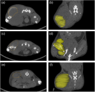
NanoPartz fluorophore labeled gold nanoparticles have solved this problem. Reference (1) explored the detectability of Nanopartz fluorescently labeled gold nanoparticles under micro-CT and optical projection tomography (OPT) in agarose phantoms and a murine melanoma tumor model with success. The distribution of the gold nanoparticles appeared to be contained and isolated to the tumor. Alignment of micro-CT specimen scans with the OPT scans was possible which highlights the potential use of fluorescently labeled gold nanoparticles for imaging murine melanoma tumors using micro-CT and OPT.
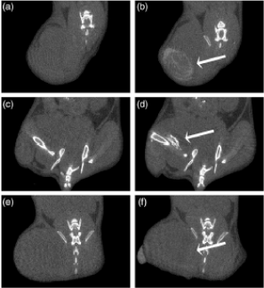
Images of Nanopartz in vivo gold nanorods providing contrast for in vivo micro-CT scan at 50 μm resolution.(a), (c), and (e) pre-contrast and (b), (d), and (f) post-contrast injection of 20 μl of gold nanorod solution of mouse #7 [(a) and (b)], #9 [(c) and (d)], and #5 [(e) and (f)]. Arrows point to tumor. From (1).
What is Micro-CT?
Micro-CT is a high-resolution imaging technique that allows for the detailed visualization of small objects, tissues, and materials in three dimensions. It uses X-rays to generate cross-sectional images, which are then reconstructed into a 3D model. Micro-CT is particularly valuable in fields such as biomedical research, materials science, and industrial inspection.
The Role of NanoPartz Gold Nanoparticles in Micro-CT
1. Enhanced Contrast
NanoPartz gold nanoparticles are ideal contrast agents for micro-CT due to their high atomic number and excellent X-ray attenuation properties. When introduced into a sample, these nanoparticles significantly enhance the contrast in the resulting images, allowing for the clear differentiation of structures that may otherwise be difficult to visualize.
2. Targeted Imaging
NanoPartz gold nanoparticles can be functionalized with specific biomolecules, enabling targeted imaging of particular tissues, cells, or molecular markers. This targeting capability is crucial for studying complex biological processes, such as tumor development, tissue regeneration, or the distribution of drugs within an organism.
3. Quantitative Analysis
The strong contrast provided by NanoPartz gold nanoparticles facilitates precise quantitative analysis in micro-CT imaging. Researchers can accurately measure the concentration and distribution of nanoparticles within a sample, providing valuable data for studies in pharmacokinetics, toxicology, and materials science.
4. Applications in Biomedical Research
In biomedical research, NanoPartz gold nanoparticles are used to enhance the imaging of soft tissues, blood vessels, and other structures in small animal models. They allow for the detailed study of disease progression, treatment efficacy, and the effects of therapeutic interventions.
5. Materials Science Applications
In materials science, these nanoparticles are utilized to improve the visualization of internal structures within composites, polymers, and other materials. Micro-CT imaging with NanoPartz gold nanoparticles enables the study of porosity, phase distribution, and the integrity of materials at the microscale.
Why Choose NanoPartz Gold Nanoparticles?
- High Purity and Consistency: NanoPartz gold nanoparticles are manufactured to the highest standards, ensuring consistent quality and performance in your imaging studies.
- Customizable Solutions: Whether you need nanoparticles with specific functionalizations or particular sizes, NanoPartz can provide tailored solutions to meet your research needs.
- Proven Results: Researchers worldwide trust NanoPartz gold nanoparticles to deliver reliable, high-contrast imaging in micro-CT studies.
References
1. Kozomara, S., & Ford, N. L. (2020). Detectability of fluorescent gold nanoparticles under micro-CT and optical projection tomography imaging. Journal of Medical Imaging, 7(2), 026002. https://doi.org/10.1117/1.JMI.7.2.026002
Conclusion
NanoPartz gold nanoparticles are revolutionizing micro-CT imaging by providing superior contrast, targeted imaging capabilities, and precise quantitative analysis. Whether you're working in biomedical research, materials science, or industrial applications, these nanoparticles will elevate the quality and depth of your imaging results.
Go here to purchase Nanopartz Fluorophore Labeled Gold Nanoparticles as used in the Kozomara and Ford article
Go here to purchase Nanopartz Gold Nanoparticles for in vitro applications
Go here to purchase Nanopartz Gold Nanoparticles for in vivo applications